Brain research to benefit from new cell-marking technique
Neurology News & Neuroscience News from Medical News Today
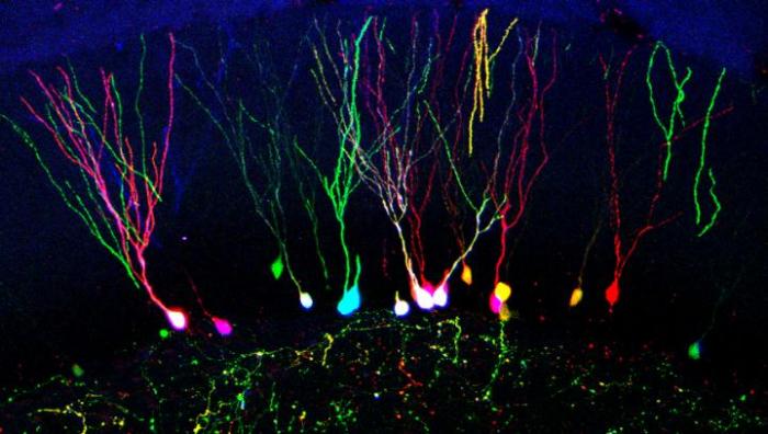
Original Article: http://www.medicalnewstoday.com/articles/287434.php
(1 of 3)
Patients with osteoporotic acute thoracic and lumbar vertebral body fractures were randomly assigned to either kyphoplasty or vertebroplasty with 12- and 24-month posttreatment fracture incidence as the primary end point. Kyphoplasty and vertebroplasty had similar long-term improvement in pain and disability with comparable safety profiles and few device-related complications. Procedure duration was shorter with vertebroplasty. Kyphoplasty had fewer cement leakages and a trend toward longer fracture-free survival.

BACKGROUND AND PURPOSE
Several trials have compared vertebral augmentation with nonsurgical treatment for vertebral compression fractures. This trial compares the efficacy and safety of balloon kyphoplasty and vertebroplasty.
MATERIALS AND METHODS
Patients with osteoporosis with 1–3 acute fractures (T5–L5) were randomized and treated with kyphoplasty (n = 191) or vertebroplasty (n = 190) and were not blinded to the treatment assignment. Twelve- and 24-month subsequent radiographic fracture incidence was the primary end point. Due to low enrollment and early withdrawals, the study was terminated with 404/1234 (32.7%) patients enrolled.
RESULTS
The average age of patients was 75.6 years (77.4% female). Mean procedure duration was longer for kyphoplasty (40.0 versus 31.8 minutes, P < .001). At 12 months, 7.8% fewer patients with kyphoplasty (50/140 versus 57/131) had subsequent radiographic fracture, and there were 8.6% fewer at 24 months (54/110 versus 64/111). The results were not statistically significant (P > .21). When we used time to event for new clinical fractures, kyphoplasty approached statistical significance in longer fracture-free survival (Wilcoxon, P = .0596). Similar pain and function improvements were observed. CT demonstrated lower cement extravasation for kyphoplasty (157/214 versus 164/201 levels treated, P = .047). For kyphoplasty versus vertebroplasty, common adverse events within 30 postoperative days were procedural pain (12/191, 9/190), back pain (14/191, 28/190), and new vertebral fractures (9/191, 17/190); similar 2-year occurrence of device-related cement embolism (1/191, 1/190), procedural pain (3/191, 3/190), back pain (2/191, 3/190), and new vertebral fracture (2/191, 2/190) was observed.
CONCLUSIONS
Kyphoplasty and vertebroplasty had similar long-term improvement in pain and disability with similar safety profiles and few device-related complications. Procedure duration was shorter with vertebroplasty. Kyphoplasty had fewer cement leakages and a trend toward longer fracture-free survival.
The post A Randomized Trial Comparing Balloon Kyphoplasty and Vertebroplasty for Vertebral Compression Fractures due to Osteoporosis appeared first on AJNR Blog.
 BACKGROUND: Several studies report early results of endoscopic endonasal transsphenoidal surgery; however, none discuss long-term outcome measures such as tumor recurrence rates and the need for additional surgical procedures. OBJECTIVE: To discuss the long-term outcomes after endoscopic endonasal transsphenoidal surgery for nonfunctioning pituitary macroadenomas. METHODS: This is a retrospective study. Patients were included only if they had at least 5 years of clinical and imaging follow-up after surgery. RESULTS: Eighty patients met the study criteria. Grossly complete resection was achieved in 71% of patients. Knosp grade 0 to 2 tumors and tumor with volumes <10 cm3 were significantly more likely to have received a grossly complete resection. There were 7 recurrences (12%) in patients who had received grossly complete resections, with a mean time to recurrence of 53 months. Among the 23 patients who had subtotal resections, 11 (61%) progressed radiographically, and 3 (17%) had symptomatic progression. Knosp score, surgical and radiographic evidence of invasion, and preoperative visual deficits were predictive of recurrence in a univariate analysis, but Knosp grade was the only independent predictor in a multivariate analysis. Kaplan-Meier analysis projected a 10-year progression-free survival rate of 80% and 21% for patients with gross total resections and subtotal resections, respectively. CONCLUSION: At the long-term follow-up, 12% of patients had recurrent tumors after grossly complete resection. Recurrent or residual tumors were treated with either repeat surgery or Gamma Knife radiosurgery. Rates of complete resection, postoperative surgical and endocrinological complications, and additional surgical procedures are similar to previously published reports after microscopic transsphenoidal surgery. ABBREVIATION: ETSS, endoscopic transsphenoidal surgeryBACKGROUND: Several studies report early results of endoscopic endonasal transsphenoidal surgery; however, none discuss long-term outcome measures such as tumor recurrence rates and the need for additional surgical procedures. OBJECTIVE: To discuss the long-term outcomes after endoscopic endonasal transsphenoidal surgery for nonfunctioning pituitary macroadenomas. METHODS: This is a retrospective study. Patients were included only if they had at least 5 years of clinical and imaging follow-up after surgery. RESULTS: Eighty patients met the study criteria. Grossly complete resection was achieved in 71% of patients. Knosp grade 0 to 2 tumors and tumor with volumes <10 cm3 were significantly more likely to have received a grossly complete resection. There were 7 recurrences (12%) in patients who had received grossly complete resections, with a mean time to recurrence of 53 months. Among the 23 patients who had subtotal resections, 11 (61%) progressed radiographically, and 3 (17%) had symptomatic progression. Knosp score, surgical and radiographic evidence of invasion, and preoperative visual deficits were predictive of recurrence in a univariate analysis, but Knosp grade was the only independent predictor in a multivariate analysis. Kaplan-Meier analysis projected a 10-year progression-free survival rate of 80% and 21% for patients with gross total resections and subtotal resections, respectively. CONCLUSION: At the long-term follow-up, 12% of patients had recurrent tumors after grossly complete resection. Recurrent or residual tumors were treated with either repeat surgery or Gamma Knife radiosurgery. Rates of complete resection, postoperative surgical and endocrinological complications, and additional surgical procedures are similar to previously published reports after microscopic transsphenoidal surgery. ABBREVIATION: ETSS, endoscopic transsphenoidal surgery
 BACKGROUND: Optimizing liposomal vehicles for targeted delivery to the brain has important implications for the treatment of brain tumors. The promise of efficient, brain-specific delivery of chemotherapeutic compounds via liposomal vehicles has yet to be achieved in clinical practice. Intra-arterial injection of specially designed liposomes may facilitate efficient delivery to the brain and to gliomas. OBJECTIVE: To test the hypothesis that cationic liposomes may be effectively delivered to both normal and glioma-bearing brain tissue utilizing a strategy of intra-arterial injection during transient cerebral hypoperfusion. METHODS: Cationic, anionic, and neutral liposomes were separately injected via the internal carotid artery of healthy rats during transient cerebral hypoperfusion. Rats bearing C6 gliomas were similarly injected with cationic liposomes. Liposomes were loaded with DilC18(5) dye whose concentrations can be measured by light absorbance and fluorescence methods. RESULTS: After intra-arterial injection, a robust uptake of cationic in comparison with anionic and neutral liposomes into brain parenchyma was observed by diffuse reflectance spectroscopy. Postmortem multispectral fluorescence imaging revealed that liposomal cationic charge was associated with more efficient delivery to the brain. Cationic liposomes were also readily observed within glioma tissue after intra-arterial injection. However, over time, cationic liposomes were retained longer and at higher concentrations in the surrounding, peritumoral brain than in the tumor core. CONCLUSION: This study demonstrates the feasibility of cationic liposome delivery to brain and glioma tissue after intra-arterial injection. Highly cationic liposomes directly delivered to the brain via an intracarotid route may represent an effective method for delivering antiglioma agents. ABBREVIATIONS: Chol, cholesterol DiD, DilC18(5) DMPC, dimyristoylphosphatidylcholine DOTAP, dioleoyl-trimethylammonium-propane IA, intra-arterial MCA, middle cerebral artery OP, optical pharmacokinetic TCH, transient cerebral hypoperfusion
Early neurological deterioration (END) following ischaemic stroke is a serious event with manageable causes in only a fraction of patients. The incidence, causes and predictors of END occurring within 24 h of acute ischaemic stroke (END24) have not been systematically reviewed. We systematically reviewed Medline and Embase from January 1990 to April 2013 for all studies on END24 following acute ischaemic stroke (<8 h from onset). We recorded the incidence and presumed causes of and factors associated with END24. Thirty-six studies were included. Depending on the definition used, the incidence of END24 markedly varied among studies. Using the most widely used change in National Institutes of Health Stroke Scale ≥4 definition, the pooled incidence was 13.8% following thrombolysis, ascribed to intracranial haemorrhage and malignant oedema each in ~20% of these. As other mechanisms were rarely reported, in the majority no clear cause was identified. Few data on END24 occurring in non-thrombolysed patients were available. Across thrombolysed and non-thrombolysed samples, the strongest and most consistent admission predictors were hyperglycaemia, no prior aspirin use, prior transient ischaemic attacks, proximal arterial occlusion and presence of early CT changes, and the most consistent 24 h follow-up associated factors were no recanalisation/reocclusion, large infarcts and intracranial haemorrhage. Finally, END24 was strongly predictive of poor outcome. The above findings are discussed with emphasis on END without a clear mechanism. Data on incidence and predictors of the latter subtype is scarce, and future studies using systematic imaging protocols should address its underlying pathophysiology. This may in turn lead to rational preventative and therapeutic measures for this ominous event.
(3 of 3)
The authors describe the imaging and pathology results of 47 patients who underwent surgery for incidentally found thyroid nodules. All patients had biopsies before surgery but only 4% of these showed benign processes. Surgery eventually demonstrated that 51% of nodules were benign and when malignant the most common histology was papillary type.
BACKGROUND AND PURPOSE
Incidental thyroid nodules are commonly seen on imaging, and their work-up can ultimately lead to surgery. We describe characteristics and pathology results of imaging-detected incidental thyroid nodules that underwent surgery.
MATERIALS AND METHODS
A retrospective review was performed of 303 patients who underwent thyroid surgery over a 1-year period to identify patients who presented with incidental thyroid nodules on imaging. Medical records were reviewed for the types of imaging studies that led to detection, nodule characteristics, and surgical pathology.
RESULTS
Of 303 patients, 208 patients (69%) had surgery for thyroid nodules. Forty-seven of 208 patients (23%) had incidental thyroid nodules detected on imaging. The most common technique leading to detection was CT (47%). All patients underwent biopsy before surgery. The cytology results were nondiagnostic (6%), benign (4%), atypia of undetermined significance or follicular neoplasm of undetermined significance (23%), follicular neoplasm or suspicious for follicular neoplasm (19%), suspicious for malignancy (17%), and diagnostic of malignancy (30%). Surgical pathology was benign in 24 of 47 (51%) cases of incidental thyroid nodules. In the 23 incidental cancers, the most common histologic type was papillary (87%), the mean size was 1.4 cm, and nodal metastases were present in 7 of 23 cases (30%). No incidental cancers on imaging had distant metastases.
CONCLUSIONS
Imaging-detected incidental thyroid nodules led to nearly one-fourth of surgeries for thyroid nodules, and almost half were initially detected on CT. Despite indeterminate or suspicious cytology results that lead to surgery, more than half were benign on final pathology. Guidelines for work-up of incidental thyroid nodules detected on CT could help reduce unnecessary investigations and surgery.
The post Imaging-Detected Incidental Thyroid Nodules that Undergo Surgery: A Single-Center Experience Over 1 Year appeared first on AJNR Blog.

A 61-year-old man presented with a 4-month history of progressive vision loss and paresthesia between the right L2-L4 dermatome. Funduscopy revealed severe bilateral papilledema (figure 1). Head CT showed mild hydrocephalus. CSF from a lumbar puncture at the L4-L5 level was xanthochromic with an elevated protein level (4,350 mg/dL). Lumbar spine MRI demonstrated a large intradural tumor at the L3-L4 level (figure 2), which was pathologically identified as schwannoma. Surgical removal resolved all symptoms. Papilledema is a rare complication induced by marked elevation of CSF protein, which may disturb CSF absorption and result in high intracranial pressure.1,2
